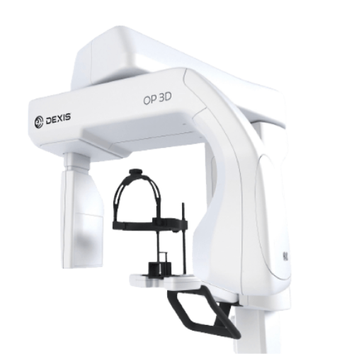
DEXIS OP 3D

By DEXIS

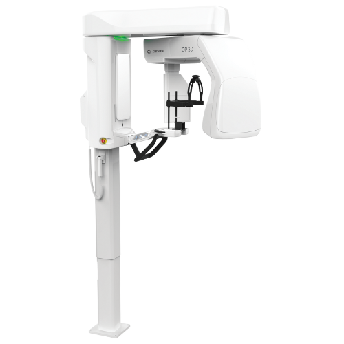
The DEXIS OP 3D is a unit designed for advanced dental imaging needs. It is a complete X-ray platform that provides easy-to-use features throughout the entire dental imaging workflow.

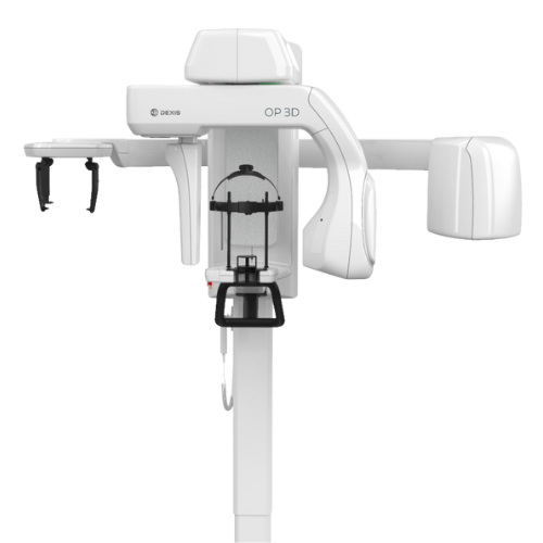
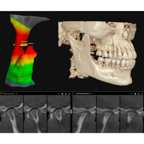
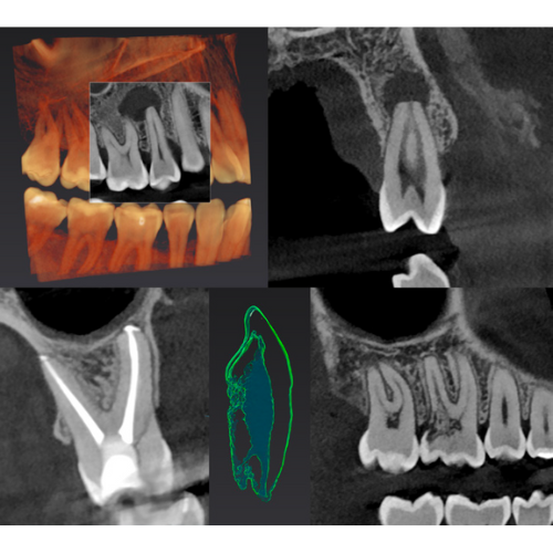
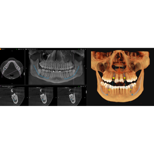
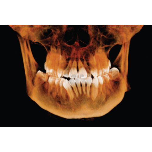
The DEXIS OP 3D offers a CBCT field of view from 5x5 cm up to 9x11 cm with an optional upgrade to 9x14 cm, covering a range of applications from endodontics to TMJ analysis. Designed to expand its capabilities as you practice grows, the DEXIS OP 3D offers an optional cephalometric attachment upgrade.


LEADING THE WAY THROUGH THE DECADES

For more than 60 years, the name ORTHOPANTOMOGRAPH system has stood for ultimate reliability and clinically correct maxillofacial imaging.

The OP 3D system respects the legacy, yet renews the meaning of image quality, efficiency and ease-of-use.

SELECTABLE FIELD OF VIEW (FOV) SIZES
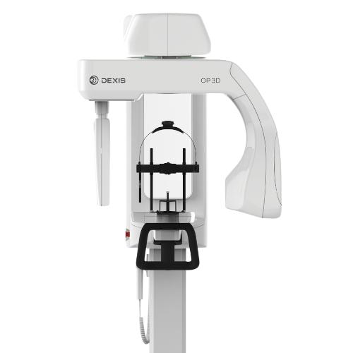
One size does not have to fit all. The OP 3D system offers efficient tools for optimizing the patient dose with its ability to allow the clinician to select the best resolution, FOV size, and region of interest.

To provide the highest level of image quality, the Metal Artifact Reduction (MAR) is readily activated with all the FOV sizes of OP 3D. The system’s graphical user interface and patient positioning system make capturing images easy while its HD image resolution with MAR ensures clear radiographs at a low radiation dose.

FULLY UPGRADEABLE

The OP 3D panoramic unit is completely upgradeable. Choose the addition of cephalometric imaging, or
completely upgrade and choose to add 3D imaging to your practice for even more diagnostic options.

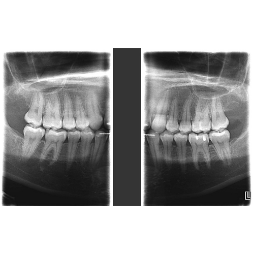
DEDICATED 2D PANORAMIC

Standard and pediatric panoramic images, along with bitewing and lateral TMJ projections cover all the extraoral imaging needs of a busy practice.

9-Second scan time: The standard panoramic program provides a clear definition of the dental anatomy, including TMJs—in only 9 seconds.

Bitewing-like view is a quick and easy alternative to intraoral bitewing imaging.

DESIGNED FOR EFFICIENCY

Every feature of the OP 3D system is designed to
increase practice efficiency. Preparing the unit for a
scan is fast with an intuitive patient positioning system
and graphical user-interface. Imaging protocols are
optimized for practice workflows

The desired imaging area can be selected
intuitively with the ORTHOselect user interface.
Selections can be made as individual teeth, an
entire upper or lower jaw, or TMJ. The optimum
field-of-view (FOV) is set automatically based on
the selection.